X-ray manual : U.S. Army . <*5 lit w Fig. 3. Position for first exposure.. Fig. 4. Position for second exposure.200 HEAD EXAMINATIONS 201 moved from the front plane of the
€ 24.50 · 4.8 (742) · Auf Lager
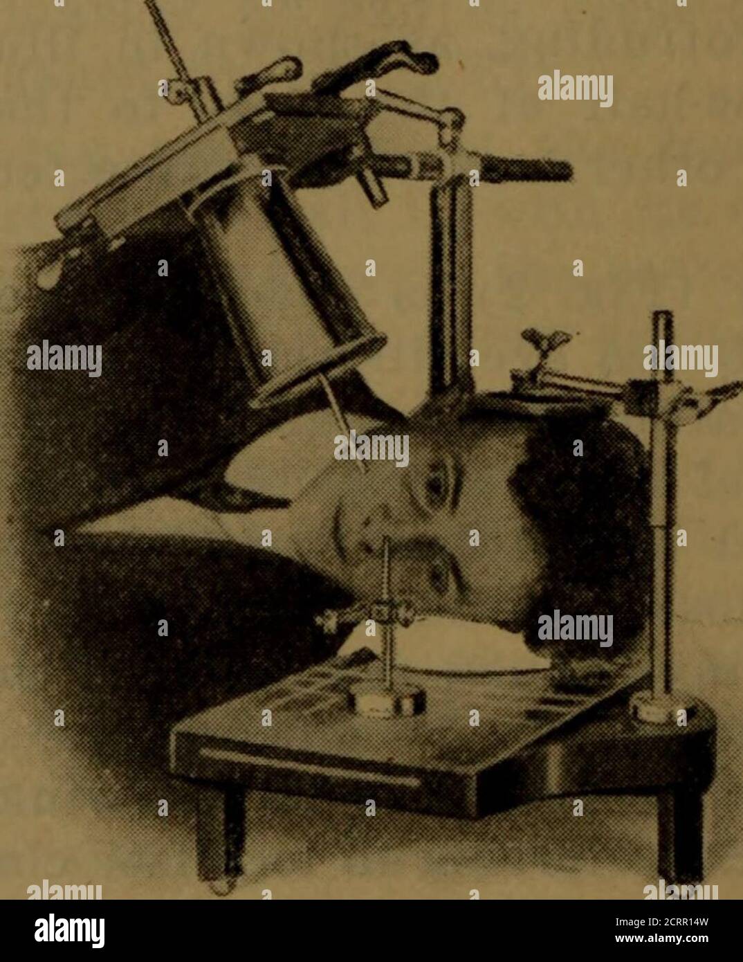
Download this stock image: . X-ray manual : U.S. Army . <*5 lit w Fig. 3. Position for first exposure.. Fig. 4. Position for second exposure.200 HEAD EXAMINATIONS 201 moved from the front plane of the cornea, and it shouldalso be borne in mind that the front of the cornea is 10millimeters in front of the shadow of the indicator-ball, asshown in your negatives. The tube is now centered overthe localizing ball and cone so that the shadows of thetwo will coincide (Fig. 3). Some object, such as a candle or a piece of whitepaper, that can be readily seen by the patient, should beplaced in alignment with the sights of the - 2CRR14W from Alamy's library of millions of high resolution stock photos, illustrations and vectors.

Preview Cambridge International AS and A Level Physics Workbook by Cambridge International Education - Issuu

Zubrick organic chemistry laboratory survival manual 2e hq by Dr.No. nofunclub - Issuu

Guidelines for the Management of TDTs (3rd ed.)- English by Thalassaemia International Federation (TIF) - Issuu

A comparative study of collimation in bedside chest radiography for preterm infants in two teaching hospitals - ScienceDirect

RBCP v35n3 - Inglês by RBCP - Issuu

Principles of Naval Weapons Systems by Francisco Azevedo - Issuu
![]()
A) Red square: Recommendations for chest x-ray boundaries; Yellow

Aviation visual Perception by batdelger - Issuu

2022 CPO Manual by thePHTA - Issuu

Electronic collimation and radiation protection in paediatric digital radiography: revival of the silver lining, Insights into Imaging
Diffi hi-res stock photography and images - Page 13 - Alamy

unified facilities criteria (ufc)

ICR 11.1 by Radcliffe Cardiology - Issuu

Catalogo Completo by Hensistemas Hensistemas - Issuu